What Does a Pinched Nerve Look Like on an MRI?
If you’re experiencing numbness, tingling, or pain in your neck, back, arms, or legs, it may be due to a pinched nerve. A pinched nerve occurs when pressure is applied to a nerve by bones, cartilage, muscles, or tendons, disrupting the nerve and causing discomfort. Some common conditions associated with a pinched nerve are herniated disks, bone spurs, and carpal tunnel syndrome.
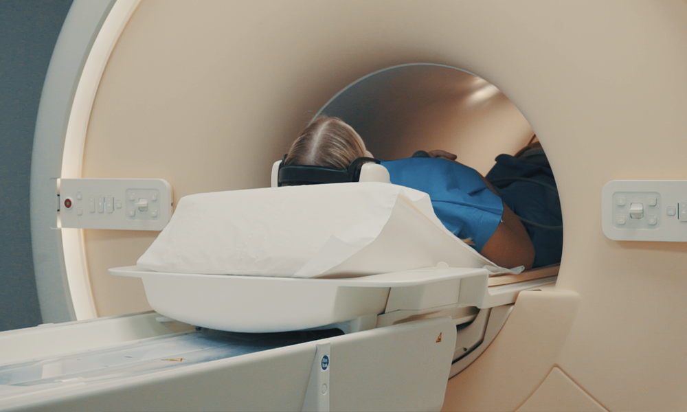
An MRI (Magnetic Resonance Imaging) scan is a medical imaging technique that uses a combination of computer-generated radio waves and a magnetic field to create detailed images of the body’s organs and tissues. It’s often used to identify the root cause of a patient’s discomfort and can be an effective way to diagnose a pinched nerve. An MRI can provide doctors with clear, detailed images of the spine from different angles (sagittal and axial), allowing them to view the vertebrae and discs closely and identify any abnormalities.
While a neurological examination can diagnose nerve damage, an MRI scan can pinpoint it more specifically. It’s important to get tested if your symptoms worsen to avoid any permanent nerve damage.
Treatment for a pinched nerve will depend on the severity of the pain and the results of the MRI scan. In many cases, doctors will recommend resting and avoiding activities that flare up the affected area. They may also recommend wearing a brace to immobilize the area. Other treatment options include physical therapy, medication, and surgery.
It’s worth noting that an MRI can only show nerve damage at the specific moment it is conducted. It cannot explicitly show if the nerve was more severely pinched a few weeks prior or how exactly the nerve is being pressed at the time of examination. Additionally, while a severe case of a bulging disc can cause permanent nerve damage, an MRI cannot always accurately predict the likelihood of this occurring.
In conclusion, a pinched nerve is a common condition that occurs when pressure is applied to a nerve, disrupting its function and causing discomfort. An MRI (Magnetic Resonance Imaging) scan is an effective way to diagnose a pinched nerve, providing clear, detailed images of the spine to help identify any abnormalities. While a neurological examination can diagnose nerve damage, an MRI scan can pinpoint it more specifically. Treatment for a pinched nerve will depend on the severity of the pain and the results of the MRI scan and may include rest, wearing a brace, physical therapy, medication, or surgery. It’s important to get tested if your symptoms worsen to avoid any permanent nerve damage. Overall, an MRI can be a valuable tool in the diagnosis and treatment of a pinched nerve.
Ready to get started?
Here’s what you’ll need to schedule an appointment
1. Imaging referral / prescription
2. Your contact information
3. Insurance OR card information


